
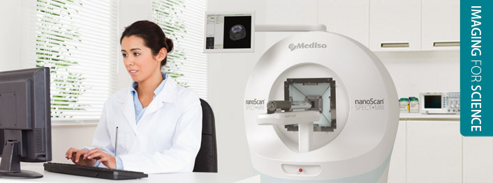
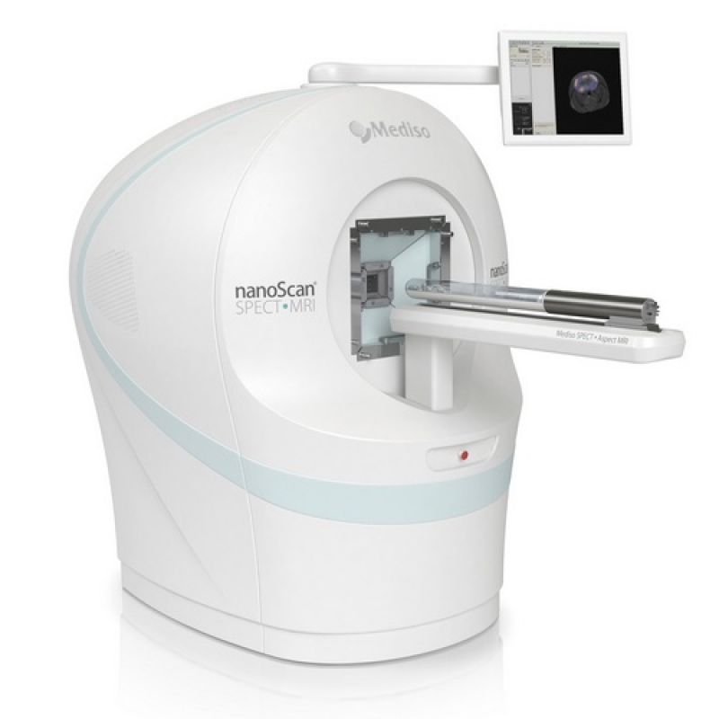
nanoScan SPET/MRI
SPECT/MRI Pre-Clinical Imaging System
First integrated in-line preclinical whole-body SPECT/MRI
275 µm SPECT and 100 µm MRI* spatial resolution
Absolute quantification accuracy over 97%
Easy to learn, easy to use in vivo multimodality imager

NANOSCAN SPECT/MRI
Integrated whole-body preclinical SPECT/MRI system for in-line multi-modality small animal imaging
Mediso pioneered integrated SPECT and MRI imaging technologies in preclinical small animal studies. SPECT/MRI represents a completely unique molecular imaging approach which allows hybrid imaging using biomarkers labeled with a wide range of radionuclides as well as single, dual- or even multi-isotope studies. Unlike PET, SPECT provides the ability to detect signals of different energies from multiple radionuclide-labeled biomarkers for fully customized and application-specific multifunctional imaging. SPECT/MRI has a variety of potential applications, including but not limited to imaging for cancer, cardiovascular and neurological diseases, thyroid and other endocrine disorders, trauma, inflammation and infection.
SPECT has no physicial limits in resolution. The imaging chain design of the nanoScan SPECT component is perfectly optimized to unleash the full power of the multi-pinhole, multi-focus and multi-size apertures. By combining the power of SPECT with state-of-the-art compact MRI technology, the nanoScan SPECT/MRI offers exceptional and unique possibilities for true translational studies. In most of the SPECT applications, soft tissue details and high contrast are desirable elements which complement SPECT dynamic, functional read-outs. The compact system is easy to install and doesn not require investment for site preparation. The plug-and-work imager is easily operated and maintained by life scientists without prior MRI experience.
SPECT/MR system includes the elimination of the radiation dose associated with CT and the much lower cost of building the technology compared to PET/MR.
Summary of Key Benefits
-
Mediso's continuous leadership in preclinical SPECT technology; based on decades of experience
- Non-multiplexed M3pinhole™ apertures, up to 100 pinholes; easily replaceable; whole-body mouse and rat
- Exceptional performance: Up to 10,000 cps/MBq sensitivity @ sub-millimeter resolution / Up to 0.275 mm resolution
- Ultra-fast 4D/5D list mode dynamic imaging by static or rotational modes with stationary and helical scans
- The ONLY small animal SPECT system to apply both attenuation and scatter correction - especially important for quantitative low-energy isotope studies
- The ONLY small animal SPECT system that can image Mice and Monkeys(*) (* with parallel hole collimators)
-
No compromise in the imaging performance: the negligible fringe field of the permanent magnet ensures the unique high performance of the SPECT subsystem
- 275 µm spatial resolution with Tera-Tomo™ 3D SPECT reconstruction engine - on the fly Monte Carlo based running on multiple GPUs
- Absolute quantification accuracy over 97%
- True stationary data collection which allows fast dynamic analysis with SPECT
- Multiple isotope imaging (unlimited number of energy windows)
-
Versatile soft tissue contrast for the best Volume of Interest definitions
- Determine the volume of tumors: for characterizing the size, vascularization and heterogeneity
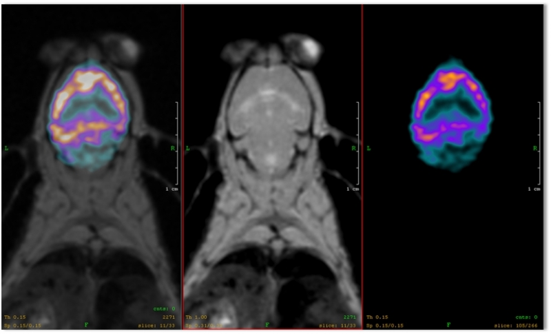
- Detailed structural information in the brain revealing exact distribution of neurological SPECT tracers
-
Compact and easy-to-use design
- does not require special training/education,
- there is no need for long calibrations after start-up
- there is no waiting time between SPECT and MRI acquisitions
- quick and easy installation
Summary of Key Benefits for high-performance, compact MRI subsystem
No active fringe magnetic field - The SPECT/MRI can be placed anywhere in a research facility (in a lab, even next to other important imaging systems)
Virtually maintenance-free
No dedicated facilities or shielded rooms, no isolation from other metal objects, simple siting and no dedicated power supplies or cooling required
Easy-to-use software, simple workflow and pre-programmed sequences - lab technician with no prior MRI or imaging experience to quickly master the platform and perform highly effective MR-based imaging experiments
The magnet operates at a 1 Tesla field strength. This is the most suitable field strength and frequency in which to perform gadolinium-based contrast agent imaging due to the nature of the relaxivity curves of such agents
Silent operation by whispering gradients – animals can be kept in the same room and depth of anesthesia can be kept at a minimum
Image resolution 100 μm in-vivo (ex-vivo spatial resolution <100 µm)
Advantages of the Compact and Integrated System
- No loss of information due to reorientation
- Attenuation & Scatter Correction based on MRI data
- Common gantry and single image coordinate system for both modalities
- Common integrated acquisition software
- Integrated and optimized SPECT/MRI acquisition protocols
- SPECT acquisition and reconstruction range selection based on MRI images
- Whole-body MRI imaging with up to 280 mm extended axial field of view
- Unequivocally co-registered MRI volume as anatomical reference for SPECT studies
- ECG and Respiratory gated imaging for both SPECT and MRI modalities
Summary of Key Benefits for SPECT
- Keeping the development in one hand: Complete detection chain redesigned
- Mediso Ultra High Performance (UHP) Detector
- More than doubled crystal volume: 50% larger detector area & 50% thicker crystal (compared to NanoSPECT)
- Fully digital premium electronics: digital 1 ADC/PMT non-Anger detector electronics
- 4 NaI(Tl) single 28x28cm crystal detectors; crystal thickness 9.5mm; detector FOV 27x27cm
- Number of photomultiplier tubes: 144 (for 4 heads)
- Fully digital: digital 1 ADC/PMT non-Anger detector electronics
- Mediso multi-focal multi-size multiple pinhole apertures (M3-pinhole™)
- Rectangular apertures have variable aperture size in one plate
- Multifocal geometry
- Non-overlapping projections
- Select the appropriate aperture for the application: whole-body mouse, whole-body rat, ultra-high sensitivity or resolution apertures
- Fast and easy replacement of apertures
- Mediso Ultra High Performance (UHP) Detector
Why Integrated?
Would you buy stand-alone SPECT and CT systems? Why would an integrated SPECT/MRI be different?
Want to achieve reproducible, sub-millimetre co-registered fusion of your modalities?
Due to the high level of integration of the nanoScan SPECT/MRI, there is no loss of information caused by reorientation and co-registration of images acquired on standalone systems.
This design allows accurate attenuation and scatter correction based on MRI datasets which is not possible with standalone or separated systems
(1) due to the inevitable misalignment of the object caused by manual positioning and
(2) due to lack of integration of SPECT reconstruction and MRI data acquisition.
Automatic co-registration is ensured by single, integrated acquisition software used by the nanoScan SPECT/MRI imager. Moreover precise positioning and automatic gantry movements enable multiple bed position MRI acquisition covering the entire body length of a mouse and rat.
The temporal correlation is quasi-simultaneous: users do not need to spend time with moving the animal from one imager to the other or even moving it from one room to another. Switching between SPECT and MRI acquisitions has never been so quick and easy before.
Considering the above mentioned advantages of the integrated nanoScan SPECT/MRI system, it is not surprising that throughput is high: acquiring a SPECT/MRI image will not take longer than a SPECT/CT with a dedicated SPECT/CT imager.
nanoScan SPECT/MRI uses the permanent magnet technology of Aspect Imaging which does not require any cooling (ie. No cryogens or helium), plumbing, or auxiliary power supplies. These features significantly reduce operational costs, require far less maintenance and allow quick and easy installation with minimal demand on the laboratory. It has small footprint and due to the negligible fringe field, any other laboratory equipment can operate right next to it without further RF or magnetic shielding. It is even compatible with any laboratory and personal electronics equipment such as mobile phones and laptops.

Ürün hakkında güncel bilgiler için satış temsilcinizle irtibat kurunuz.
Bildirimde bulunmaksızın veri değiştirme hakkı firmada saklıdır.